Published: 09/09/2025
ORIGINAL ARTICLES
Purpose of the study. Was to investigate the prognostic significance of the AKT/mTOR signaling pathway components, transcription, and growth factors in the development of skin melanoma recurrence.
Patients and methods. The study included 48 patients with a verified diagnosis of skin melanoma. All patients had a negative status for the BRAFV600E mutation. The study material consisted of samples of tumor and unchanged skin located at a distance of at least 1 cm from the tumor border, obtained during surgical treatment, which were frozen and stored at a temperature of –80 °C after sampling. Gene expression was assessed by real-time PCR. The status of the BRAF gene was assessed using allele-specific PCR. Statistical processing was carried out using the Statistica 12.0 software package.
Results. Predicting the course of cancer is an important task in practical oncology. The unfavorable outcome of melanomas is largely due to the tendency to relapses and metastasis in both the short and long-term follow-up period. While analyzing the study results, we noted the association of AKT/mTOR signaling pathway components with the development of skin melanoma recurrence and disease progression. It was revealed that the expression level of AKT, c-RAF, GSK‑3β and VEGFR2 is associated with an unfavorable outcome of the disease. A logistic regression model is presented that can accurately predict the risk of recurrence, taking into account the expression of protein kinase mTOR and the size of the tumor (T). The lack of significant indicators related to the biological features of the skin tumor does not allow to increase the effectiveness of treatment of such patients. Therefore, the attention of researchers is focused on finding optimal models for predicting the risk of an unfavorable outcome of the disease.
Conclusion. Molecular markers were identified that make it possible to predict an unfavorable outcome of the disease during the study. A logistic model based on the expression of the key mTOR kinase and the size of the T has been developed, which makes it possible to assess the risk of disease recurrence and change treatment tactics in a timely manner.
Purpose of the study. The objective of this study is to evaluate the clinical effectiveness of activation xenon-oxygen therapy (XOT) in correcting neuropsychological and adaptive status in patients diagnosed with hormone-dependent breast cancer who have undergone total ovariectomy.
Patients and methods. We analyzed data on the intensity of clinical symptoms, the dynamics and intensity of adaptive responses (AR) with calculation of the anti-stress/stress ratio, as well as the bioelectric brain activity of 36 young patients diagnosed with hormone-dependent breast cancer, presenting with clinical signs of post-mastectomy syndrome (PMES) and early signs of post-ovariectomy syndrome (POES).
Results. In determining the distribution of stress and anti-stress reactions, a postoperative phase transition from a physiological to a pathological state was reliably established. In the postoperative period following ovariectomy, normal reaction types were observed in 11.3 % of cases. The predominant response was the stress reaction. Analysis of AR structure in patients with PMES and POES manifestations demonstrated a clear advantage for XOT. During rehabilitation, patients in the non-XOT group did not achieve full restoration of AR structure to baseline values, with the stress response persisting in 58.7 % of cases. In contrast, the XOT group demonstrated anti-stress reactions, with no stress reactions detected. Furthermore, analysis of the bioelectric activity of the brain in patients after two hormone-reducing surgeries revealed significant alterations in the spectral power of the electroencephalogram (EEG). These changes were indicative of a balanced state of excitation and inhibition, suggesting an equilibrium in neural processes underlying cognitive function.
Conclusion. Xenon, as a biologically active factor, functions as a catalyst for complex functional transformations within the body's regulatory systems. Xenon-based therapy induces a substantial reduction in stress reactions, highlighting the pronounced biotropic effect of xenon in restoring the adaptive status of the female body.
Purpose of the study. The objective of this study was to evaluate cell cycle indices in tumor cells and conditionally intact intestinal tissue (resection line) in male and female patients with colorectal cancer (CRC).
Patients and methods. Cell cycle phases were analyzed in 36 male and 36 female patients with CRC involving the rectum, and with right-sided or left-sided tumor localization. The mean age was 66 years. The degree of tumor differentiation in all cases corresponded to G2. None of the patients had received neoadjuvant treatment before surgery. In 10 % of tumor homogenates and resection line samples, cell cycle phases were determined using an ADAMII LS fluorescent cell analyzer (Korea). For cell cycle analysis, propidium iodide (PI), specially prepared for the ADAMII LS, was used. This reagent mixture contained PI and RNaseA, and cells were stained directly without an additional fixation step. The instrument’s high sensitivity enabled precise discrimination of cells in the G0/G1 phase (resting cells [G0] and early G1), S phase, and G2/M phase. Statistical analysis was performed using Statistica 10.0 software.
Results. The proportions of viable and dead cells in the samples were generally comparable. In men, viable cells ranged from 56.6 % to 73.6 %, and dead cells from 26.4 % to 43.4 %. In women, viable cells ranged from 60.8 % to 77.3 %, and dead cells from 22.7 % to 39.2 %. In men, tumor samples from left-sided CRC contained predominantly S and G2 phase cells, whereas in right-sided CRC and rectal tumors, the majority of cells were in the G1 phase. In women with left-sided CRC, tumor samples showed the highest proportion of cells in the G1 phase, while samples from right-sided CRC and rectal tumors contained predominantly S phase cells.
Conclusion. The identified cell cycle characteristics and tumor cell death patterns in CRC patients, depending on sex and tumor localization, reflect the proliferative status of the tissue. These findings may provide a basis for personalized recommendations on the use of antitumor agents targeting cells in specific cell cycle phases.
Purpose of the study. To perform a preliminary assessment of local control after two-stage staged radiosurgery in patients with metastatic brain lesions.
Patients and methods. For staged radiosurgery, large lesions measuring ≥ 3 cm in the largest dimension were selected. The regimen consisted of delivering 12 Gy in a single fraction at the first stage and 14 Gy in a single fraction at the second stage, with a 14‑day interval between the stages. If additional smaller lesions were present, they were irradiated simultaneously using the standard SRS technique in a single fraction with a dose per fraction (DPF) of 18–24 Gy. The prospective analysis included 32 patients of both sexes aged 34 to 76 years (mean age 57 ± 3.3 years) with brain metastatic lesions ≥ 3 cm in the largest dimension, or located in close proximity to critical brain structures, who underwent a two-stage course of staged radiosurgery at the National Medical Research Centre for Oncology.
Results. The evaluation of target lesion volumes was based on brain MRI performed before treatment, prior to the second stage, and one month after completion of treatment. At the one-month follow-up after the treatment course, local control was achieved in the vast majority of clinical cases. Sixteen lesions demonstrated a volume reduction of more than 70 % from baseline, eleven showed a reduction of more than 50 %, eight lesions exhibited a decrease of less than 50 %, and one lesion demonstrated a negative response.
Conclusion. Two-stage staged radiosurgery for brain metastases demonstrated satisfactory local control in patients with various primary tumor sites. The positive dynamics observed at this stage suggest the potential for favorable long-term outcomes.
Purpose of the study. To investigate the feasibility of applying a method of visual diagnosis of basal cell carcinoma (BCC) under ultraviolet (UV) light using Photoditazine for determining tumor margins in clinical practice.
Patients and Methods. The study was conducted at the Department of Reconstructive and Plastic Surgery and Oncology, National Medical Research Center for Oncology. Sixty patients (men and women) with cytologically verified stage I–II BCC were included. Among them, 30 patients (15 men and 15 women) presented with a superficial growth pattern, and 30 patients (15 men and 15 women) with a solid growth pattern. In all cases, Photoditazine gel-penetrant was applied topically to the tumor surface and the surrounding area (1.5–2 cm) for 30 minutes. The gel was then removed with a gauze swab moistened with distilled water. Under UV light in a dark room, a characteristic fuchsia fluorescence of the tumor and a pale or dark-red halo around the lesion were observed. This enabled assessment of the tumor spread into adjacent tissues that initially appeared visually unaffected.
Results. The study yielded the following findings: in 33 patients, the peritumoral area showed no fluorescence, whereas in 27 patients, zones of enhanced fluorescence ("hot spots") were detected around the tumor. These areas exhibited a round or irregular shape and were characterized by an intense dark-red fluorescence surrounding the tumor focus. Extensive "hot spots" indicated active adsorption of the photosensitizer in the area, which may result from altered hormonal and metabolic properties of the peritumoral skin. In cases where extensive peritumoral fluorescence was identified, the area should be included within the resection field as a potentially high-risk zone for subsequent recurrence.
Conclusion. The proposed method is simple to use, does not require expensive equipment or systemic administration of the photosensitizer, and therefore avoids patient inconvenience and precautionary restrictions. Detection of peritumoral fluorescence enables accurate determination of true tumor margins and facilitates subsequent surgical excision with an optimal margin from the visible edge of the lesion. These advantages support the recommendation of this method for broad implementation in routine practice of specialized medical institutions.
Purpose of the study. To analyze the correlation between the proliferative activity of atypical cells in triple-negative breast cancer (BC) and patient age.
Patients and methods. The study included 80 women with a newly diagnosed BC, in whom a triple-negative surrogate molecular genetic subtype was identified based on histological and immunohistochemical examination with assessment of hormone receptor expression and Ki‑67. Statistical analysis was performed using Statistica 13.5.0.17 software (TIBCO Software Inc.). The Shapiro-Wilk test was applied to assess the normality of the distribution. Correlation between variables was evaluated using the Kendall tau rank correlation coefficient.
Results. The median age of patients was 53.9 years (95 % confidence interval [CI]: 50.0–57.8) (Fig. 3). Among them, 65 % of patients were younger than 50 years and 35 % were older. The median proliferative activity, assessed by Ki‑67 expression according to the St. Gallen Consensus (2009), was 76.4 % (95 % CI: 73.28–79.66). However, this indicator varied depending on patient age. The analysis revealed a correlation between Ki‑67 expression level and patient age, with a Kendall tau coefficient of –0.449 (p < 0.05), corresponding to a weak-to-moderate negative association.
Conclusion. The analysis showed that the degree of Ki‑67 expression correlates with the age at breast cancer onset. Thus, there is a tendency toward higher proliferative activity of cancer cells in younger patients.
REVIEW
This article provides an overview of current approaches to cancer immunotherapy, with an emphasis on the role of dendritic cells (DCs), lymph nodes (LNs), and innovative methods of vaccine delivery. Immunotherapy using DC-based vaccines represents a promising direction, capable of stimulating a specific immune response against tumor cells and forming long-term immune memory. Tumor-draining lymph nodes (TDLNs) play a key role in immune activation, as they are the sites where dendritic cells present tumor antigens and activate T-cells. In cancer, unlike viral infections, CD8+ T-cell activation occurs in two stages, and the effectiveness of this process depends on signals from the tumor microenvironment, which explains why the immune response to cancer is often weak.
The article also discusses modern strategies for delivering vaccines to lymph nodes, including the use of nanoparticles, bioorthogonal reactions, and photothermally induced materials. These approaches help overcome the "granularity paradox", associated with the need to balance vaccine size for LN penetration and uptake by immune cells. The prospects of adoptive cell therapy using T-cells from TDLNs, as well as the role of exosomes and whole-cell tumor antigens in the development of effective vaccines, are also considered. Combination strategies, such as the use of vaccines together with checkpoint inhibitors (e. g., anti-PD1), demonstrate potential for enhancing antitumor immunity.
The further advancement of cancer immunotherapy requires the integration of new knowledge about the biology of dendritic cells, modern methods of cell engineering, and nanotechnology to create personalized and effective antitumor vaccines.
ЮБИЛЕЙ
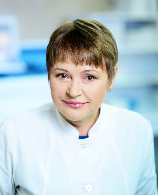
Elena M. Frantsiyants, Doctor of Biological Sciences, Professor, Deputy General Director for Scientific Work at the National Medical Research Center for Oncology, and Honored Healthcare Worker of the Russian Federation, celebrate her 70th birthday on September 23, 2025.
Dr. Frantsiyants is a distinguished scientist, pathophysiologist, and biochemist whose contributions to the understanding of the fundamental mechanisms of tumor development are invaluable. Born in Taganrog into a family of educators, she chose her path early, dedicating her life to science. After graduating from the Faculty of Biology and Soil Science at Rostov State University in 1978, Dr. Frantsiyants began her professional career at the Central Research Laboratory of Rostov State Medical University. In 1985, she joined the Rostov Scientific Research Oncological Institute, where she progressed from senior researcher to head of the Biochemical Laboratory (1990–2000), and later to head of the Laboratory for the Study of the Pathogenesis of Malignant Tumors (2014–2017). Since 2017, she has served as Deputy General Director for Science.
Her scientific interests center on the pathogenesis of tumors, particularly the role of antioxidant systems in carcinogenesis and tumor growth. Her pioneering research has uncovered novel aspects of metabolic restructuring that contribute to tumor initiation and progression. These findings laid the groundwork for the introduction of biochemical markers into clinical practice for predicting disease course and evaluating antitumor treatment efficacy. Moreover, she developed a set of biochemical techniques for the individualization of targeted cancer therapies. Dr. Frantsiyants has also made significant contributions to understanding hormonal imbalances and mediator dysfunctions in the development of malignancies.
Currently, she leads research at the National Medical Research Center for Oncology focusing on mitochondrial dysfunction in malignant tumor growth, and the experimental development of targeted therapies and dendritic cell vaccines.
Her extensive body of work includes over 1,000 scientific publications, 205 patents, and 8 monographs, such as Lipid Peroxidation in the Pathogenesis of Tumor Disease (1995), Complex Treatment of Primary Malignant Glial Tumors of the Cerebral Hemispheres (2014), Cytological, Morphological and Immunohistochemical Diagnostics of Central Nervous System Tumors (2015), Pathogenetic Aspects of Metastatic Liver Damage: An Experimental Study (2022), and Neuroendocrine and Metabolic Aspects of Melanoma Pathogenesis: An Experimental and Clinical Study (2023).
Over her more than 40-year tenure at the National Medical Research Center of Oncology, Dr. Frantsiyants has emerged as a leading expert in basic oncology research. She serves as a scientific consultant, supervises graduate and doctoral candidates (7 Ph.D. and 7 doctoral dissertations), and holds positions as Deputy Chair of the Academic Council and member of the Dissertation Council at the National Medical Research Center for Oncology.
Professor Frantsiyants is not only a brilliant and passionate scientist endowed with deep knowledge, intellectual rigor, and analytical prowess, but also a person of warmth, grace, and the rare ability to balance scientific achievement with a fulfilling family life.
The staff of the National Medical Research Center for Oncology and the editorial board of the South Russian Journal of Cancer extend their heartfelt congratulations to Dr. Frantsiyants on her anniversary! We wish you continued health, prosperity, new scientific accomplishments, and boundless energy in the pursuit of your goals. May your research continue to serve society and earn the recognition it so richly deserves. You have rightfully earned deep respect and admiration from your colleagues and students alike!
On August 31, 2025, the scientific community and colleagues sincerely congratulate Oleg Ivanovich Kit, Academician of the Russian Academy of Sciences, Doctor of Medical Sciences, Professor, and General Director of the National Medical Research Centre for Oncology, on his 55th anniversary!
Dr. Oleg I. Kit is an outstanding oncologist and a recognized leader in the field of healthcare organization and higher medical education. His 30-year scientific and practical activity at the National Medical Research Centre for Oncology is reflected in more than 1,000 publications, including 21 monographs, 15 study guides, and 236 patents of the Russian Federation.
Dr. Kit is the Editor-in-Chief of the scientific and practical journal South Russian Journal of Cancer, which is reviewed by the Higher Attestation Commission. He is also a member of the editorial boards of the journals Voprosy Onkologii, Russian Journal of Oncology, Oncology. P. A. Herzen Journal of Oncology, Problems of Hematology/Oncology and Immunopathology in Pediatrics, Biomedicine, Science in the South Russia, and Cardiometry.
Under the leadership of Dr. Kit, over a 15-year period, an authoritative scientific school in the field of oncology and oncosurgery was formed, oriented toward the development and implementation of innovative surgical approaches and treatment methods based on an in-depth study of the molecular-genetic, pathogenetic, and immunological mechanisms of malignant neoplasms. He is the initiator of the introduction of organ-preserving, reconstructive, and minimally invasive operations, as well as the improvement of surgical techniques aimed at the prevention of postoperative complications.
Dr. Kit made a significant contribution to the creation of modern infrastructure for oncological research, including the organization of a unified pathological and anatomical center and the establishment of a comprehensive depository of tumor samples and DNA and RNA isolated from them, which made it possible to conduct large-scale genetic research. Under his leadership, studies are being conducted on the molecular profiling of pancreatic neuroendocrine tumors, the identification of prognostic markers of cardiotoxicity, the determination of molecular-genetic predictors of the course of gliomas, as well as preclinical trials of new antitumor agents.
For the development and implementation of an interdisciplinary strategy in the treatment of colorectal cancer, in 2016 he was awarded the Prize of the Government of the Russian Federation in the field of science and technology and was given the title of "Laureate of the Prize of the Government of the Russian Federation in the field of science and technology".
As the Chief Freelance Oncologist of the Ministry of Health of the Russian Federation in the Southern Federal District, he devotes great attention to raising the professional level of physicians. For the development of oncological care and the improvement of cancer treatment in the Southern Federal District, in 2020 Dr. Kit was awarded the N. N. Trapeznikov Medal "For Contribution to the Development of Oncology Service".
Since 2015, Dr. Kit has been Head of the Department of Oncology of Rostov State Medical University.
Dr. Kit is the Chairman of the Academic Council and the Dissertation Council for the defense of doctoral and candidate dissertations at the National Medical Research Centre. Under his supervision, 21 doctoral and 23 candidate dissertations have been successfully defended.
The large-scale work of O. I. Kit in the field of education, science, practical medicine, organizational and public activity has been recognized by numerous state, ministerial, and public awards, including the badge of distinеction "Excellence in Healthcare", the Medal of the Order “For Merit to the Fatherland" II degree, the Medal "For Merit to Russian Healthcare", the Medal of the Order "For Merit to the Rostov Region", and a Letter of Gratitude from the President of the Russian Federation.
His high professionalism, sense of duty, attentive and benevolent attitude toward people have earned him authority and respect among the scientific community, colleagues, students, and patients.
The staff of the National Medical Research Centre for Oncology and the editorial board of the South Russian Journal of Cancer sincerely congratulate Dr. Kit on his anniversary! We wish You good health, well-being, inexhaustible energy, new scientific achievements, and continued success in your responsible and multifaceted work! May your knowledge and experience continue to serve the advancement of Russian science and healthcare.


